
How MRIs are Changing How Society Looks at your Brain

Have you had an experience in an MRI scanner? Chances are you have or know someone who has. Magnetic Resonance Imaging produces detailed pictures of soft tissue in the human body – but, unlike many other techniques, it does not require any radiation. A doctor from the 1950s would be shocked at the images obtained with non-invasive modern MRI scanners of structures inside the body – they’ve come a long way since the technology at that time. 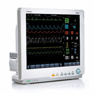 These remarkable machines are doing surprising things, all at the hands of signal processing.Have you had an experience in an MRI scanner? Chances are you have or know someone who has. Magnetic Resonance Imaging produces detailed pictures of soft tissue in the human body – but, unlike many other techniques, it does not require any radiation. A doctor from the 1950s would be shocked at the images obtained with non-invasive modern MRI scanners of structures inside the body – they’ve come a long way since the technology at that time. These remarkable machines are doing surprising things, all at the hands of signal processing.
If you have ever visited a hospital, you’ve probably noticed the many waveforms that are displayed near the patient’s bedside. What you may not have realized is the degree to which sophisticated signal processing is going on in the hospital room: pulse rate and oxygen level provided by a simple-looking device on the finger, a cardiac signal from a few sensors on the chest, and even more information from other sensors. Signal processing can give doctors a breadth of information, all from just a few sensors.
For example, a baseball pitcher who comes to the hospital for a scan of his/her shoulder won’t know that the images collected from the scan use a form of one of the most common signal processing algorithms, known as the FFT (fast Fourier transform algorithm). But that pitcher will be desperately interested in whether a part of their shoulder shows something sinister in the resulting image, no matter how subtle that feature might be.
Besides the striking images it produces, a new development is that there are ways to use MRI now to ascertain the health of microscopic cellular structures in the brain. Structures such as the brain’s white matter, including the dendrites connecting one neuron to others. The white matter is mainly responsible for making the brain’s many connections.
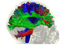
By following the diffusion pathways of water molecules in the brain, MRI now allows the construction of images showing the complex network of nerve fibers connecting different brain areas. This process is known as fiber tractography.Image 2 includes the outline of a brain and the pathways, the white matter fiber tracts, for some of the connections between neurons. Researchers can now make maps of the connections from one part of the brain to another.
 |
These advances stem from the theory of Diffusion MRI. Diffusion Tensor Imaging and its more advanced approach called Diffusion Kurtosis Imaging (DKI) provide very detailed, accurate and quantifiable information about the white matter in the brain.
These new methods employ MRI scans and DKI in ways that may eventually lead to biomarkers related to many disorders, including Alzheimer’s Disease, Multiple Sclerosis, Alcoholism, Autism, ADHD, and more. Similarly, “… use of Diffusion Kurtosis Imaging can be expected to improve the ability to diagnosis Parkinson’s disease.3”
This novel imaging modality could, for example, help predict the onset of disease long before the first clinical symptoms appear. If this method provides the information as anticipated, Alzheimer’s Disease could receive a much earlier diagnosis.
MRI would simply not exist without advances in signal processing and it’s helping it expand in amazing ways. Electronic radio-frequency technology is responsible for generating, shaping, and amplifying the electrical currents required to produce the magnetic field for the scanner. And now, it’s responsible for exploring areas of your body you could have never expected. Just imagine what’s yet to come!
Further reading:
- “Mapping the Brain with Diffusional Kurtosis,” in MUSC Health Progressnotes, Features, Summer 2015. Medical University of South Carolina, online at www.muschealth.org.
- “MR Diffusion Tensor Imaging: A Window into White Matter Integrity of the Working Brain,” by S. Chanraud, N. Zahr, E. V. Sullivan, and A. Pfefferbaum. Neuropsychol Rev. pp.209–225, 2010.
- “Diffusional kurtosis imaging of cingulate fibers in Parkinson disease: Comparison with conventional diffusion tensor imaging,” by K. Kamagata et al, Magnetic Resonance Imaging, pp. 1501–1506. 2013.
Dale Mugler is chief technology officer at Ocius Technologies, LLC in Akron, Ohio.