The Next Frontier of Medical Imaging: Where Signal Processing Meets AI An interview with Suthambhara Nagaraj, Principal AI Architect for MRI at Philips
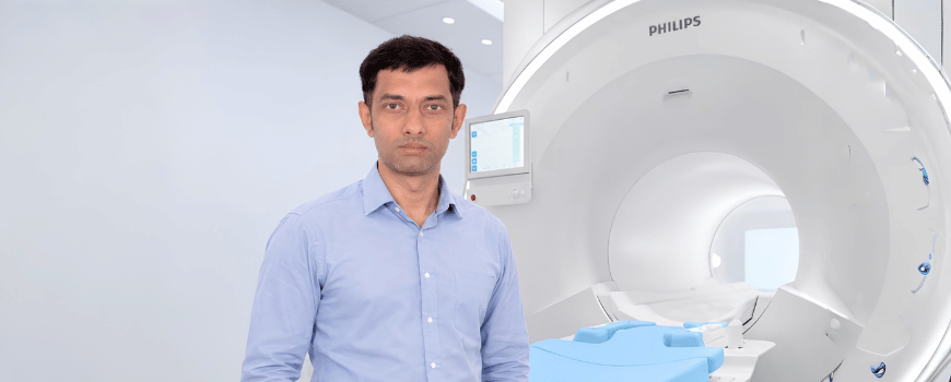
By Mariya Doneva, Editor of the IEEE SPS Industry Signals Newsletter
Mariya: Dear Suthu, thank you for joining me for this conversation on signal processing and AI in medical imaging. Public discussions are often dominated by bold predictions that AI will one day replace radiologists. However, I believe our readers are equally interested in understanding the opportunities and challenges that AI presents from the perspective of the medical imaging industry, where new technologies must ultimately deliver clinical value and improve patient care.
Let us begin by exploring the relationship between signal processing and AI in this context.
AI is currently transforming healthcare, but AI ultimately depends on high-quality signals and data. How do you see the relationship between classical signal processing and modern AI evolving over the coming years in the field of medical imaging?
Suthu: When a patient enters an imaging room, the scanner does not see anatomy directly. It captures faint, indirect, and imperfect signals. Whether in MRI, ultrasound, CT, PET, or physiological monitoring, those measurements must first be transformed into images, maps, or quantitative biomarkers before a clinician can act on them. That transformation is enabled by signal processing: modelling the measurement, managing noise and artifacts, estimating uncertainty, and producing information that has clinical meaning.
AI extends this foundation; it does not replace it. The most useful medical AI systems are not simply trained to recognize patterns in pixels. They are designed with an understanding of how the signal was acquired, how the image was formed, and what kinds of noise or variability are expected. This distinction matters. An image reconstruction algorithm should not merely produce a sharp or visually convincing image. It must preserve clinically relevant information, perform predictably across scanners, hospitals, and patient populations, and reveal its limitations. An image that appears realistic but invents detail not supported by the acquired data is not an improvement; it can be misleading.
Mariya: This is indeed a very important point, especially in the context of medical data. In your view, how important are signal quality, data fidelity, and physics-based modeling in ensuring trustworthy clinical AI? How is this considered in the context of regulatory approval of medical devices?
Suthu: This becomes even more important as generative AI enters medical imaging. Generative models can create a highly plausible output, but plausibility is not the same as fidelity to the measured signal for a particular patient. Signal processing gives AI essential guardrails: grounding outputs in measured data, incorporating known physics and noise behavior, and using anatomical or temporal constraints where appropriate. The future of medical imaging is therefore not a competition of classical signal processing versus AI, but their integration. Signal processing brings physical grounding, robustness, and interpretability, while AI learns complex patterns from large and diverse datasets.
Trust, however, must extend beyond the architecture. It begins with the data. Medical datasets may underrepresent particular patient groups, disease presentations, scanners, sites, or acquisition protocols. A model can look successful on average while being unreliable in the settings where it is most needed. We must therefore understand data provenance, assess its representativeness, and test deliberately across clinically relevant variation. Data quality deserves the same discipline: labels may be incomplete or subjective, image quality can vary, and curation can introduce errors. We need to characterize these sources of uncertainty, define acceptance criteria, and maintain traceability from data to performance claims.
Ultimately, clinical trust is earned through evidence, not only benchmark accuracy, but also an understanding of failure modes, robustness in real-world use, workflow usability, and the consequences of error. This is a continuous engineering discipline, not a one-time validation exercise.
From a regulatory perspective, the term trustworthy AI is translated into concrete requirements for safety, effectiveness, risk management, transparency, robustness and post market monitoring. The FDA has established a framework for AI -enables medical devices and more than 1600 devices have been approved to date.
Mariya: Medical imaging has relatively high adoption of AI including methods enabling faster MR scans, dose reduction in CT, improved image quality, as well as image processing. What other areas are on the horizon and how can they impact patient care, e.g. quantitative biomarkers, or autonomous workflows?
Suthu: That rigor can unlock a much larger benefit. The next frontier is moving from AI that improves an individual image to AI that improves the entire care pathway. Healthcare demand is rising faster than the available clinical workforce, so the real opportunity is to use technology to give clinicians more time for decisions and patient interactions where their expertise matters most.
In imaging, this means increasingly autonomous and standardized workflows across modalities. AI can help select and tailor protocols, guide patient positioning, plan examinations, optimize acquisition in real time, detect motion or incomplete coverage early, and ensure that image quality is sufficiently consistent for diagnosis. This could allow one technologist to remotely supervise several systems, while an on-site technologist or a nurse can focus their attention on patient comfort, safety, and the exceptions that genuinely require human judgement.
A second important shift is from images as primarily visual information to images as quantitative measurements. Reliable biomarkers such as organ volumes, tissue characteristics, perfusion quantification, fat and iron measurements, lesion burden, or cardiac function can make disease assessment more objective and reproducible. When tracked over time, they can support earlier detection of change, more personalized treatment decisions, and clearer communication across sites and care teams.
Finally, AI can connect data acquisition, image reconstruction, image processing, quantification, and reporting into a more coherent workflow. The aim is not to replace radiologists, technologists, or other healthcare professionals. It is to extend their capacity: reduce avoidable process variability and repetitive work, improve access to high-quality imaging, and help more patients receive timely, confident diagnosis and treatment.
Mariya: Suthu, thank you for sharing your insights. Our discussion highlights that the future of medical imaging is not a choice between signal processing and AI, but a powerful combination of both. While AI is enabling new possibilities, staying grounded in the principles of signal processing remains essential to ensure data fidelity, robustness, and trust in the results.
Ultimately, the success of AI in radiology and in healthcare in general will be measured by its ability to help clinicians work more effectively and improve patient care. It will be exciting to see how this field continues to evolve and shape the future of medical imaging.
Thank you again for sharing your perspective with the readers of IEEE SPS Industry Signals.
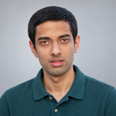 Dr. Suthambhara Nagaraj is an AI Architect in Philips’ MR business. He received his PhD in Computer Science from the Indian Institute of Science. His work spans magnetic resonance image reconstruction, with particular experience in AI-based reconstruction methods that improve image quality, acquisition efficiency, and robustness. In his current role, he helps shape the development and translation of trustworthy AI technologies for clinical MR imaging.

Dr. Mariya Doneva is a Senior Scientist at Philips Innovative Technologies. She received her PhD in Physics from the University of Lübeck. Her research focuses on quantitative MRI, accelerated image acquisition, and advanced image reconstruction, including compressed sensing and deep learning. Her innovations have enabled substantial reductions in MRI examination times and have been translated