If you’ve ever been to the emergency room due to an injury, chances are you’ve had a routine encounter with what is in fact a wonder of the modern age: Magnetic Resonance Imaging, or MRI. Advancements in medical imaging allow us to study parts of the body, including the brain, to an extent unprecedented in the history of medical science. But how are MRI images formed safely – non-invasively and without radiation? The short answer is that they rely on physics and signal processing. And signal processing engineers are developing radical new ways to study the human brain and body safely through medical imaging technology.
How Does MRI Work?
MRI leverages magnetic fields so powerful that they are 60,000 times stronger than the standard refrigerator magnet. This magnetic field targets hydrogen atom protons, making each proton align with or against the field. Repeated radiofrequency current pulses are introduced. When a pulse is turned on, it knocks the protons out of alignment, and when it is turned off, the protons gradually realign. As the radiofrequency pulse is turned on and off repeatedly, millions of radio signals are generated. Gradients in the magnetic field provide this data to different pixel locations. This output forms what is called the K-space, which are the spatial frequency components of the MRI image. Signal processing is key to this entire process. To create the MRI image, we apply a dominant signal processing algorithm known as the Inverse of the Fast Fourier Transform on the K-space data. The Fast Fourier Transform, or “FFT,” is one of the most versatile tools for scientists and engineers and identifies the frequencies in a signal.
Overcoming Limitations in Medical Imaging Technology
But signal processing engineers are exploring ways to optimize the image construction process. Most MRIs take at least an hour or more to produce a perfect image. This has two negative effects: logistical strain on the healthcare facility (scanning a limited number of patients), and patient discomfort and claustrophobia. (Anyone who has had to wait inside an MRI machine knows how frustrating it can be!) There is a need for faster turnaround. To create images more quickly, certain frequency K-space data is often omitted or truncated, applying the Inverse Transform only on the remaining data. But this isn’t an ideal solution; it results in the field of view being smaller than the body part being imaged (a process called “aliasing artifacts”), including the Gibbs phenomenon, which is a ringing that arises from truncating the K-space frequencies. 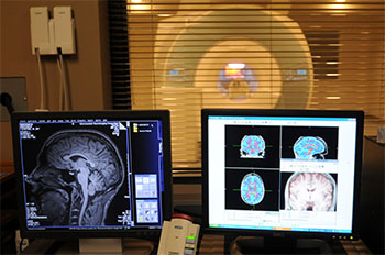
So how do we get better images without waiting longer? Signal processing engineers are developing techniques to reduce these problems, including Compressed Sensing (CS). Compressed Sensing uses sophisticated signal processing techniques to construct an image from partial K-space samples – resulting in faster image creation, but without the Gibbs ringing. These techniques are still evolving and are an area of opportunity for signal processing engineers to collaborate with doctors and technicians to disrupt the medical imaging industry. In fact, Facebook’s Artificial Intelligence Research (FAIR) group and the NYU School of Medicine have partnered to explore how artificial intelligence can use parallel imaging to generate scans faster.
Exploring the Brain: Radical New Innovations in MRI
Neurological disorders will place an increasing burden on health care systems over the coming decades, and technology is needed to help scientists and doctors better understand how these diseases form and progress. Engineers are leveraging signal processing in new, innovative MRI techniques that study functions of the brain, which remains mysterious to medical science. As these techniques evolve, engineers can use them to:
- Develop “wiring maps” of neural connections in the brain, which will aid surgeons in brain tumor removals
- Improve MR spectroscopy, another noninvasive test that measures biochemical changes in the brain, especially the presence of tumors.
- Advance MRI Diffusion Imaging methods, which are used to study Alzheimer’s, Parkinson’s and ADHD, and also predict who will benefit from a given therapy.
- Determine which parts of the brain are active when you tell a lie.
These are all radical new innovations that can substantially change the way we think about our brains and help us solve the biggest health problems impacting the world. Will you lead the next wave of signal processing that will transform the medical imaging industry? To learn more about these technologies, contact the IEEE Signal Processing Society Image, Video and Multidimensional Signal Processing Technical Committee.