- Our Story
- Publications & Resources
- Publications & Resources
- Publications
- IEEE Signal Processing Magazine
- IEEE Journal of Selected Topics in Signal Processing
- IEEE Signal Processing Letters
- IEEE Transactions on Computational Imaging
- IEEE Transactions on Image Processing
- IEEE Transactions on Information Forensics and Security
- IEEE Transactions on Multimedia
- IEEE Transactions on Signal and Information Processing over Networks
- IEEE Transactions on Signal Processing
- IEEE TCI
- IEEE TSIPN
- Data & Challenges
- Submit Manuscript
- Guidelines
- Information for Authors
- Special Issue Deadlines
- Overview Articles
- Top Accessed Articles
- SPS Newsletter
- SigPort
- SPS Resource Center
- Publications FAQ
- Blog
- News
- Dataset Papers
- Conferences & Events
- Community & Involvement
- Professional Development
- For Volunteers
- Information for Authors-OJSP
-
Home
Waveforms for Computing Over the Air: A groundbreaking approach that redefines data aggregation
Ode to Masterfully Written Textbooks: And remembering Simon Haykin [From the Editor]
Conferences Events IEEE Signal Processing Magazine IEEE SPL Article IEEE TIFS Article IEEE TMM Article IEEE TSP Article Jobs in Signal Processing Lectures Machine Learning Seasonal Schools Signal Processing News SPM Article SPS Distinguished Lectures SPS Newsletter Article SPS Webinar SPS Webinars SPS Webinar Series Webinar webinars -
Our Story
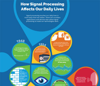
What is Signal Processing?
The technology we use, and even rely on, in our everyday lives –computers, radios, video, cell phones – is enabled by signal processing. Learn More » -
Publications & Resources
-
SPS Resources
- Signal Processing Magazine The premier publication of the society.
- SPS Newsletter Monthly updates in Signal Processing
- SPS Resource Center Online library of tutorials, lectures, and presentations.
- SigPort Online repository for reports, papers, and more.
- SPS Feed The latest news, events, and more from the world of Signal Processing.
-
SPS Resources
-
Conferences & Events
-
Community & Involvement
-
Membership
- Join SPS The IEEE Signal Processing Magazine, Conference, Discounts, Awards, Collaborations, and more!
- Chapter Locator Find your local chapter and connect with fellow industry professionals, academics and students
- Women in Signal Processing Networking and engagement opportunities for women across signal processing disciplines
- Students Scholarships, conference discounts, travel grants, SP Cup, VIP Cup, 5-MICC
- Young Professionals Career development opportunities, networking
- Get Involved
-
Technical Committees
- Applied Signal Processing Systems
- Audio and Acoustic Signal Processing
- Bio Imaging and Signal Processing
- Computational Imaging
- Image Video and Multidimensional Signal Processing
- Information Forensics and Security
- Machine Learning for Signal Processing
- Multimedia Signal Processing
- Sensor Array and Multichannel
- Signal Processing for Communication and Networking
- Signal Processing Theory and Methods
- Speech and Language Processing
- Technical Working Groups
- More TC Resources
-
Membership
-
Professional Development
-
Professional Development
- Signal Processing Mentorship Academy (SigMA) Program
- Micro Mentoring Experience Program (MiME)
- Distinguished Lecturer Program
- Distinguished Lecturers
- Distinguished Lecturer Nominations
- Past Lecturers
- Distinguished Industry Speaker Program
- Distinguished Industry Speakers
- Distinguished Industry Speaker Nominations
- Industry Resources
- IEEE Training Materials
- Jobs in Signal Processing: IEEE Job Site
-
Career Resources
- SPS Education Program Educational content in signal processing and related fields.
- Distinguished Lecturer Program Chapters have access to educators and authors in the fields of Signal Processing
- Job Opportunities Signal Processing and Technical Committee specific job opportunities
- Job Submission Form Employers may submit opportunities in the area of Signal Processing.
-
Professional Development
-
For Volunteers
-
For Board & Committee Members
- Board Agenda/Minutes* Agendas, minutes and supporting documentation for Board and Committee Members
- SPS Directory* Directory of volunteers, society and division directory for Board and Committee Members.
- Membership Development Reports* Insight into the Society’s month-over-month and year-over-year growths and declines for Board and Committee Members
-
For Board & Committee Members


Popular Pages
Today's:
- Information for Authors
- (ASRU 2025) 2025 IEEE Automatic Speech Recognition and Understanding Workshop
- IEEE Transactions on Information Forensics and Security
- IEEE Transactions on Multimedia
- Membership
- IEEE Transactions on Image Processing
- (ICME 2026) 2026 IEEE International Conference on Multimedia and Expo
- IEEE Signal Processing Letters
- Submit a Manuscript
- IEEE Journal of Selected Topics in Signal Processing
- Information for Authors-SPL
- IEEE Transactions on Audio, Speech and Language Processing
- Inside Signal Processing Newsletter
- Unified EDICS
- IEEE Transactions on Signal Processing
All time:
- Information for Authors
- Submit a Manuscript
- IEEE Transactions on Image Processing
- IEEE Transactions on Information Forensics and Security
- IEEE Transactions on Multimedia
- IEEE Transactions on Audio, Speech and Language Processing
- IEEE Signal Processing Letters
- IEEE Transactions on Signal Processing
- Conferences & Events
- IEEE Journal of Selected Topics in Signal Processing
- Information for Authors-SPL
- Conference Call for Papers
- Signal Processing 101
- IEEE Signal Processing Magazine
- Guidelines
Last viewed:
- (ASRU 2025) 2025 IEEE Automatic Speech Recognition and Understanding Workshop
- Nominations Open for IEEE Transactions on Multimedia Editor-in-Chief
- IEEE Transactions on Multimedia
- Awards & Submit Award Nomination
- 2025 IEEE International Workshop on Information Forensics and Security (WIFS)
- SPS BSI Webinar: NeuroAI: From HoloBrain to HoloGraph
- Signal Processing Cup
- IEEE Signal Processing Cup 2024
- (ICME 2026) 2026 IEEE International Conference on Multimedia and Expo
- Editorial Board
- Editorial Board
- Information for Authors
- (MLSP 2025) 2025 IEEE International Workshop on Machine Learning for Signal Processing
- Digital Edition-Content Gazette
- IEEE Signal Processing Magazine
Recent Patents in Signal Processing (June 2016) – medical imaging
You are here
Newsletter Menu
Newsletter Categories


Top Reasons to Join SPS Today!
1. IEEE Signal Processing Magazine
2. Signal Processing Digital Library*
3. Inside Signal Processing Newsletter
4. SPS Resource Center
5. Career advancement & recognition
6. Discounts on conferences and publications
7. Professional networking
8. Communities for students, young professionals, and women
9. Volunteer opportunities
10. Coming soon! PDH/CEU credits
Click here to learn more.
News and Resources for Members of the IEEE Signal Processing Society
Recent Patents in Signal Processing (June 2016) – medical imaging
For our June 2016 issue, we cover recent patents dealing with hardware and signal processing issues of medical imaging.
According to Patent no. 9,349,178, in hemodynamic determination in medical imaging, the classifier is trained from synthetic data rather than relying on training data from other patients. A computer model (in silico) may be perturbed in many different ways to generate many different examples. The flow is calculated for each resulting example. A bench model (in vitro) may similarly be altered in many different ways. The flow is measured for each resulting example. The machine-learnt classifier uses features from medical scan data for a particular patient to estimate the blood flow based on mapping of features to flow learned from the synthetic data. Perturbations or alterations may account for therapy so that the machine-trained classifier may estimate the results of therapeutically altering a patient-specific input feature. Uncertainty may be handled by training the classifier to predict a distribution of possibilities given uncertain input distribution. Combinations of one or more of uncertainty, use of synthetic training data, and therapy prediction may be provided.
Patent no. 9,349,067 introduces a method and apparatus for correcting image data from a medical imaging scan of a subject, into which subject a specified amount of an imaging substance has been introduced, a region of the image data, containing an anomalous proportion of the imaging substance introduced, is identified. For the identified region a regional value of a variable in the image data associated with the imaging substance is determined. The regional value is used to determine the proportion of the substance in the region, and the proportion is subtracted from the specified amount of the imaging substance
The claim no. D756,517 presents the ornamental design for a multi-purpose medical imaging device support apparatus.
Patent no. 9,342,922 presents and apparatus and a method of medical diagnostic imaging. The apparatus includes: an image unit for constructing volume image data by capturing images from a multiplicity of tomographic images of a sampling specimen and for constructing internal three dimensional images of the diagnosing object of the sampling specimen as seen from a viewing point; a display for displaying the three-dimensional images; an input unit for entering parameters for setting up a precutting plane at an inter-voxel image data boundary between voxel image data of the volume image data closer to the viewing point than the diagnosing object and voxel image data associated with the diagnosing object; and a control unit for controlling the structure of the three-dimensional images constructed by the image unit based on the precutting plane set up via the input unit, wherein the control unit extracts a boundary based on one of the parameters inputted to the input unit.
Patent no. 9,336,605 introduces a medical imaging system for acquiring medical image data, the medical imaging system comprising: a tissue treating system for treating a target volume; a computer system comprising a processor, wherein the computer system is adapted for controlling the medical imaging system; and a memory containing machine readable instructions. Execution of the instructions cause the processor to: acquire medical image data; reconstruct a medical image using the medical image data; receive an image segmentation seed derived from a treatment plan, and identify a treated volume in the medical image by segmenting the medical image in accordance with the image segmentation seed.
The claim no. D755,209 proposes the ornamental design for a medical imaging display screen or portion thereof with graphical user interface, as shown and described.
Patent no. 9,330,801 inntroduces a photon collimator, suitable for use in medical imaging equipment, is constructed from a block of photon-attenuating material, such as solid tungsten or molybdenum alloy that defines a plurality of integrally formed septa slats. Each slat has an elongated length dimension greater than thickness and depth dimensions, and is oriented in an opposed pattern array that is laterally spaced relative to its respective thickness dimension. An aperture channel is defined between each pair of opposed slats. Rows of integrally formed slats in one block or separately affixed blocks may be stacked on each other at skewed angles to form two-dimensional grids of apertures having polygonal cross sections. The slats may be formed by electric discharge or laser thermal ablation machining, such as by a sequential passing of an EDM wire cutting head along the pattern array, repeating sequential cutting of respective channel depth and width.
The invention no. 9,330,454 relates to a computer-implemented method of performing image quality analysis on images from an imaging examination, including: displaying an imaging examination; performing a quality review of the images; performing a quality analysis on data from the imaging examination and on the images; receiving image quality analysis from the user on the data and the images and storing the image quality analysis in at least one database; saving data from the image quality analysis on at least one key image of the images, using computer-generated standardized annotation and mark-up, or graphical data input, or speech data, using a computerized tool, and saving the data as annotated image data in the at least one database; and transferring the annotated image data to at least one quality assurance database with linking to images from the imaging examination
If you have an interesting patent to share when we next feature patents related to medical imaging, or if you are especially interested in a signal processing research field that you would want to be highlighted in this section, please send email to Csaba Benedek (benedek.csaba AT sztaki DOT mta DOT hu).
References
Number: 9,349,178
Title: Synthetic data-driven hemodynamic determination in medical imaging
Inventors: Itu; Lucian Mihai (Brasov, RO), Passerini; Tiziano (Plainsboro, NJ), Rapaka; Saikiran (Pennington, NJ), Sharma; Puneet (Monmouth Junction, NJ), Schwemmer; Chris (Forchheim, DE), Schoebinger; Max (Hirschaid, DE), Redel; Thomas (Poxdorf, DE), Comaniciu; Dorin (Princeton Junction, NJ)
Issued: May 24, 2016
Assignee: Siemens Aktiengesellschaft (Munchen, DE)
Number: 9,349,067
Title: Method and apparatus for correcting medical imaging data
Inventors: Kelly; Matthew David (Botley, GB)
Issued: May 24, 2016
Assignee: Siemens Medical Solutions USA, Inc. (Malvern, PA)
Number: D756,517
Title: Multi-purpose medical imaging device support apparatus
Inventors: Sagalovich; Boris (New York, NY), Ukrainsky; Gennady (New York, NY), Kopit; Danny (Brooklyn, NY)
Issued: May 17, 2016
Assignee: Comprehensive Telemedicine (Forest Hills, NY)
Number: 9,342,922
Title: Medical imaging apparatus and method of constructing medical images
Inventors: Nishiura; Tomofumi (Tokyo, JP)
Issued: May 17, 2016
Assignee: HITACHI MEDICAL CORPORATION (Tokyo, JP)
Number: 9,336,605
Title: Medical imaging system, computer-implemented method, and computer program product for identifying a treated region in a medical image
Inventors: Nieminen; Heikki Juhani (Helsinki, FI), Kohler; Max Oskar (Espoo, FI), Hakkinen; Marko Tapani (Espoo, FI)
Issued: May 10, 2016
Assignee: Koninklijke Philips N.V. (Eindhoven, NL)
Number: D755,209
Title: Medical imaging display screen or portion thereof with graphical user interface
Inventors: Bonnaudet; Jean-Marc (Munich, DE), Hau; Alexander (Tuttlingen, DE), Hiltl; Christoph (Bohlingen, DE), Holoien; Lee D. (Santa Barbara, CA)
Issued: May 3, 2016
Assignee: Karl Storz Imaging, Inc. (Goleta, CA)
Number: 9,330,801
Title: Method for fabricating medical imaging multilayer, multiaperture collimator
Inventors: Malmin; Ronald E. (Chicago, IL)
Issued: May 3, 2016
Assignee: Siemens Medical Solutions USA, Inc. (Malvern, PA)
Number: 9,330,454
Title: Method and apparatus for image-centric standardized tool for quality assurance analysis in medical imaging
Inventors: Reiner; Bruce (Berlin, MD)
Issued: May 3, 2016
Open Calls
Society News
- Introducing the SPS Resource Center
- 32 Signal Processing Society Members Elevated to Senior Member
- Upcoming Distinguish Lectures
- Now Live! New IEEE Signal Processing Society Website
- Call for Nominations: Chapter of the Year Award
- Call for Nominations: Awards Board Chair
- Important Message Requiring Your Vote
- SPS Job Marketplace
Conferences & Events
PhD Theses
- Simek, Kyle LouisView Profile. (The University of Arizona), “Branching Gaussian process models for computer vision” (2016)
- Briggs, GordonView Profile. (Tufts University), “Toward Dialogue and Reasoning Mechanisms to Enable More Natural and Socially-Appropriate Task-Based Human-Robot Interactions” (2016)
Technical Committee News
Publications News
Education & Resources
SPS Social Media
- IEEE SPS Facebook Page https://www.facebook.com/ieeeSPS
- IEEE SPS X Page https://x.com/IEEEsps
- IEEE SPS Instagram Page https://www.instagram.com/ieeesps/?hl=en
- IEEE SPS LinkedIn Page https://www.linkedin.com/company/ieeesps/
- IEEE SPS YouTube Channel https://www.youtube.com/ieeeSPS
IEEE SPS Educational Resources
IEEE SPS Resource Center

IEEE SPS YouTube Channel

Home | Sitemap | Contact | Accessibility | Nondiscrimination Policy | IEEE Ethics Reporting | IEEE Privacy Policy | Terms | Feedback
© Copyright 2025 IEEE - All rights reserved. Use of this website signifies your agreement to the IEEE Terms and Conditions.
A public charity, IEEE is the world's largest technical professional organization dedicated to advancing technology for the benefit of humanity.